
Antibody and Dye Based Assays

Biotium CD3e(Rabbit PAb), CF488A conjugate, 0.1mg/mL
The immunogen for this Mab is derived from the cytoplasmic domain of CD3 epsilon chain. CD3 consists of five different polypeptide chains (designated as gamma, delta, epsilon, zeta, and eta) with MW ranging from 16-28 kDa. The CD3 complex is closely associated at the lymphocyte cell surface with the T cell antigen receptor (TCR). Reportedly, CD3 complex is involved in signal transduction to the T cell interior following antigen recognition. The CD3 antigen is first detectable in early thymocytes and probably represents one of the earliest signs of commitment to the T cell lineage. In cortical thymocytes, CD3 is predominantly intra-cytoplasmic. However, in medullary thymocytes, it appears on the T cell surface. CD3 antigen is a highly specific marker for T cells, and is present in majority of T cell neoplasms.Primary antibodies are available purified, or with a selection of fluorescent CF Dyes and other labels. CF Dyes offer exceptional brightness and photostability. No

Biotium Cytokeratin, LMW(AE-1), CF488A conjugate, 0.1mg/mL
This MAb recognizes the 56.5 kDa (CK10); 50 kDa (CK14); 50 kDa (CK15); 48 kDa (CK16); 40 kDa (CK19) keratins of the acidic (Type I or LMW) subfamily. Twenty human keratins are resolved with two-dimensional gel electrophoresis into acidic (pI 6.0) subfamilies. The acidic keratins have molecular weights (MW) of 56.5, 55, 51, 50, 50', 48, 46, 45, and 40 kDa. MAb AE3 recognizes the 65-67, 64, 59, 58, 56, and 52 kDa keratins of basic subfamily. Many studies have shown the usefulness of keratins as markers in cancer research and tumor diagnosis. AE1/AE3 is a broad spectrum anti pan-keratin antibody cocktail, which differentiates epithelial tumors from non-epithelial tumors e.g. squamous vs. adenocarcinoma of the lung, liver carcinoma, breast cancer, and esophageal cancer.Primary antibodies are available purified, or with a selection of fluorescent CF Dyes and other labels. CF Dyes offer exceptional brightness and photostability. Note: Conjugates of blue fluorescent dyes like CF
Biotium Cytokeratin 8(H1), CF488A conjugate, 0.1mg/mL
Cytokeratin 8 (CK8) belongs to the type II (or B or basic) subfamily of high molecular weight cytokeratins and exists in combination with cytokeratin 18 (CK18). CK8 is primarily found in the non-squamous epithelia and is present in majority of adenocarcinomas and ductal carcinomas. It is absent in squamous cell carcinomas. Hepatocellular carcinomas are defined by the use of antibodies that recognize only cytokeratin 8 and 18. CK8 exists on several types of normal and neoplastic epithelia, including many ductal and glandular epithelia such as colon, stomach, small intestine, trachea, and esophagus as well as in transitional epithelium. Anti-CK8 does not react with skeletal muscle or nerve cells. Epithelioid sarcoma, chordoma, and adamantinoma show strong positivity corresponding to that of simple epithelia (with antibodies against CK8, CK18 and CK19). Reportedly, anti-CK8 is useful for the differentiation of lobular (ring-like, perinuclear) from ductal (peripheral-predominant)
Biotium CD56 / NCAM(NCAM1/784), CF488A conjugate, 0.1mg/mL
This MAb reacts with an extracellular domain (close to transmembrane) of CD56/NCAM. Three isoforms of neural cell adhesion molecule (NCAM) are produced by differential splicing of the RNA transcript from a single gene. The 135 kDa isoform is the basic molecule, which is glycosylated or sialylated to produce the mature species. Anti-CD56 recognizes two proteins of the neural cell adhesion molecule, the basic molecule expressed on most neuroectodermally derived tissues and neoplasms (e.g. retinoblastoma, medulloblastomas, astrocytomas, neuroblastomas, and small cell carcinomas). It is also expressed on some mesodermally derived tumors (rhabdomyosarcoma). Anti-CD56 plays an important role in the diagnosis of nodal and nasal NK/T-cell lymphomas.Primary antibodies are available purified, or with a selection of fluorescent CF Dyes and other labels. CF Dyes offer exceptional brightness and photostability. Note: Conjugates of blue fluorescent dyes like CF405S and CF405M are not
Biotium CD48(156-4H9), CF488A conjugate, 0.1mg/mL
Reacts with human CD48, a 45 kDa glycosyl phophatidyl-inositol (GPI)-anchored cell surface protein. CD48 is strongly expressed on lymphocytes and monocytes and weakly on granulocytes but is absent on platelets, fibroblasts, epithelium and endothelium. CD48 is one of marker for detecting the defects of GPI anchoring structure on the patients with paroxysmal nocturnal hemoglobulinuria (PNH) and serves as a low affinity ligand for CD2.Primary antibodies are available purified, or with a selection of fluorescent CF Dyes and other labels. CF Dyes offer exceptional brightness and photostability. Note: Conjugates of blue fluorescent dyes like CF405S and CF405M are not recommended for detecting low abundance targets, because blue dyes have lower fluorescence and can give higher non-specific background than other dye colors.
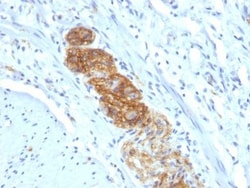
Biotium Neurofilament (NF-L)(NR-4), CF488A conjugate, 0.1mg/mL
This MAb reacts with a 68 kDa protein, identified as light sub-unit of neurofilaments (NF-L). Neurofilaments make up the main structural elements of axons and dendrites and are found in neurons, peripheral nerves, and sympathetic ganglion cells. Neurofilaments consist of three major subunits with molecular weights of 68 kDa (NF-L), 160 kDa (NF-M) and 200 kDa (NF-H). Anti-neurofilament stains a number of neural, neuroendocrine, and endocrine tumors. Neuromas, ganglioneuromas, gangliogliomas, ganglioneuroblastomas, and neuroblastomas stain positively for anti-neurofilament. Neurofilaments are also present in paragangliomas as well as adrenal and extra-adrenal pheochromocytomas. Carcinoids, neuroendocrine carcinomas of the skin, and oat cell carcinomas of the lung also express neurofilament.Primary antibodies are available purified, or with a selection of fluorescent CF Dyes and other labels. CF Dyes offer exceptional brightness and photostability. Note: Conjugates of blue fl
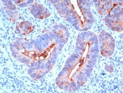
Biotium HER-2 / CD340(HRB2/258), CF488A conjugate, 0.1mg/mL
Recognizes a protein of 185 kDa, which is identified as c-erbB-2/HER-2/neu. Its epitope is localized in the extracellular domain. C-erbB-2/HER-2 is a member of the EGFR family. This MAb is specific and shows minimal cross-reaction with other members of the EGFR-family. Receptors of this family are located on the plasma membrane and consist of an extracellular ligand-binding domain that is connected to a large intracellular domain by a single transmembrane sequence. C-erbB-2/HER-2 protein is over-expressed in a variety of carcinomas especially those of breast and ovary.Primary antibodies are available purified, or with a selection of fluorescent CF Dyes and other labels. CF Dyes offer exceptional brightness and photostability. Note: Conjugates of blue fluorescent dyes like CF405S and CF405M are not recommended for detecting low abundance targets, because blue dyes have lower fluorescence and can give higher non-specific background than other dye colors.
Biotium HER-2 / CD340(HRB2/273), CF488A conjugate, 0.1mg/mL
This MAb is specific to c-erbB-2/HER-2 and shows minimal cross-reaction with other members of the family. C-erbB-2/HER-2 is a member of the EGFR family. Receptors of this family are located on the plasma membrane and consist of an extracellular ligand-binding domain that is connected to a large intracellular domain by a single transmembrane sequence. c-erbB-2/HER-2 protein is over-expressed in a variety of carcinomas especially those of breast and ovary.Primary antibodies are available purified, or with a selection of fluorescent CF Dyes and other labels. CF Dyes offer exceptional brightness and photostability. Note: Conjugates of blue fluorescent dyes like CF405S and CF405M are not recommended for detecting low abundance targets, because blue dyes have lower fluorescence and can give higher non-specific background than other dye colors.
Biotium CD66 (CEA)(C66/195), CF488A conjugate, 0.1mg/mL
This antibody recognizes proteins of 80-200 kDa, identified as different members of CEA family. CEA is synthesized during development in the fetal gut and is re-expressed in increased amounts in intestinal carcinomas and several other tumors. This MAb reacts with nonspecific cross-reacting antigen (NCA) and shows a cross-reaction with human polymorphonuclear leucocytes. It shows no reaction with a variety of normal tissues and is suitable for staining of formalin/paraffin tissues. CEA is not found in benign glands, stroma, or malignant prostatic cells. Antibody to CEA is useful in detecting early foci of gastric carcinoma and in distinguishing pulmonary adenocarcinomas (60-70% are CEA ) from pleural mesotheliomas (rarely or weakly CEA ). Anti-CEA positivity is seen in adenocarcinomas from the lung, colon, stomach, esophagus, pancreas, gallbadder, urachus, salivary gland, ovary, and endocervix.Primary antibodies are available purified, or with a selection of fluorescent CF Dy
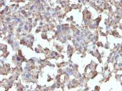
Biotium CD284 / TLR4(TLR4/230), CF488A conjugate, 0.1mg/mL
This MAb reacts with human Toll-like receptor 2 (TLR4). It is a member of the Toll-like receptor (TLR) family, which plays a fundamental role in pathogen recognition and activation of innate immunity. TLRs are highly conserved from Drosophila to humans and share structural and functional similarities. They recognize pathogen-associated molecular patterns that are expressed on infectious agents, and mediate the production of cytokines necessary for the development of effective immunity. The various TLRs exhibit different patterns of expression. This receptor has been implicated in signal transduction events induced by lipopolysaccharide (LPS) found in most gram-negative bacteria. Mutations in this gene have been associated with differences in LPS responsiveness. Multiple transcript variants encoding different isoforms have been found for this gene.Primary antibodies are available purified, or with a selection of fluorescent CF Dyes and other labels. CF Dyes offer exceptional
Biotium Progesterone(6-5E-3F), CF488A conjugate, 0.1mg/mL
This MAb is specific for progesterone. It exhibits minimal cross reactivity with related compounds in ELISA. It reacts with Progesterone-11a-HMS-BSA: 100%; 5-beta-Pregnane-3,20-dione: 48%; 5-alpha-Pregnane-3,20-dione: 26.4%; 17-alpha-Hydroxyprogesterone: 2.5%; 20-alpha-Hydroxyprogesterone: 0.04%. Progesterone is a steroid hormone synthesized from the cholesterol derivative, pregnenolone, in the cortex of the adrenal gland. Progesterone is secreted by the corpus luteum and acts to prepare the endometrium for the implantation of a fertilized egg. During pregnancy, it is secreted by the placenta to prevent spontaneous abortion and to stimulate the development of mammary tissue to produce milk. Thus, progesterone plays a central role in the reproductive events associated with the establishment and maintenance of pregnancy. Luteinized theca cells of normal ovary secrete progesterone. The determination of progesterone concentrations in the body fluids is of great value for endocrin
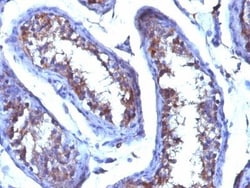
Biotium Chromogranin A(CGA/414), CF488A conjugate, 0.1mg/mL
Chromogranin A is present in neuroendocrine cells throughout the body, including the neuroendocrine cells of the large and small intestine, adrenal medulla and pancreatic islets. It is an excellent marker for carcinoid tumors, pheochromocytomas, paragangliomas, and other neuroendocrine tumors. Co-expression of chromogranin A and neuron specific enolase (NSE) is common in neuroendocrine neoplasms. Reportedly, co-expression of certain keratins and chromogranin indicates neuroendocrine lineage. The presence of strong anti-chromogranin staining and absence of anti-keratin staining should raise the possibility of paraganglioma. The co-expression of chromogranin and NSE is typical of neuroendocrine neoplasms. Most pituitary adenomas and prolactinomas readily express chromogranin.Primary antibodies are available purified, or with a selection of fluorescent CF Dyes and other labels. CF Dyes offer exceptional brightness and photostability. Note: Conjugates of blue fluorescent dyes
Biotium Chromogranin A(CHGA/413), CF488A conjugate, 0.1mg/mL
Chromogranin A is present in neuroendocrine cells throughout the body, including the neuroendocrine cells of the large and small intestine, adrenal medulla and pancreatic islets. It is an excellent marker for carcinoid tumors, pheochromocytomas, paragangliomas, and other neuroendocrine tumors. Co-expression of chromogranin A and neuron specific enolase (NSE) is common in neuroendocrine neoplasms. Reportedly, co-expression of certain keratins and chromogranin indicates neuroendocrine lineage. The presence of strong anti-chromogranin staining and absence of anti-keratin staining should raise the possibility of paraganglioma. The co-expression of chromogranin and NSE is typical of neuroendocrine neoplasms. Most pituitary adenomas and prolactinomas readily express chromogranin.Primary antibodies are available purified, or with a selection of fluorescent CF Dyes and other labels. CF Dyes offer exceptional brightness and photostability. Note: Conjugates of blue fluorescent dyes